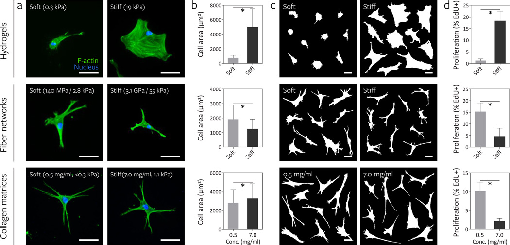
Figure 3. Increasing fiber stiffness suppresses cell spreading and proliferation.
The effect of altering material stiffness on hMSC spreading and proliferation was examined on DexMA hydrogels (top row, soft: 290 Pa, stiff: 19.1 kPa) and fiber networks (middle row, soft: 140 MPa fiber, 2.8 kPa network; stiff: 3.1 GPa fiber, 55 kPa network). Low (0.5 mg/mL, <0.3 kPa) and high (7.0 mg/mL, 1.1 kPa) concentration type I collagen matrices where bulk stiffness and adhesive ligand density increase in tandem were included for comparison (bottom row). a, Actin cytoskeletal organization of representative hMSCs 16 h after seeding, stained for F-actin (green) and cell nuclei (blue) (scale bars, 50 µm). b, Quantification of cell area; mean ± s.d., n ≥ 64 cells, * P < 0.05. c, Cell outlines of ten representative cells (scale bars, 50 µm). d, Proliferation of hMSCs over two days as determined by EdU incorporation; mean ± s.d., n ≥ 13 ROI with totals of 750–1500 cells analyzed, * P < 0.05.

