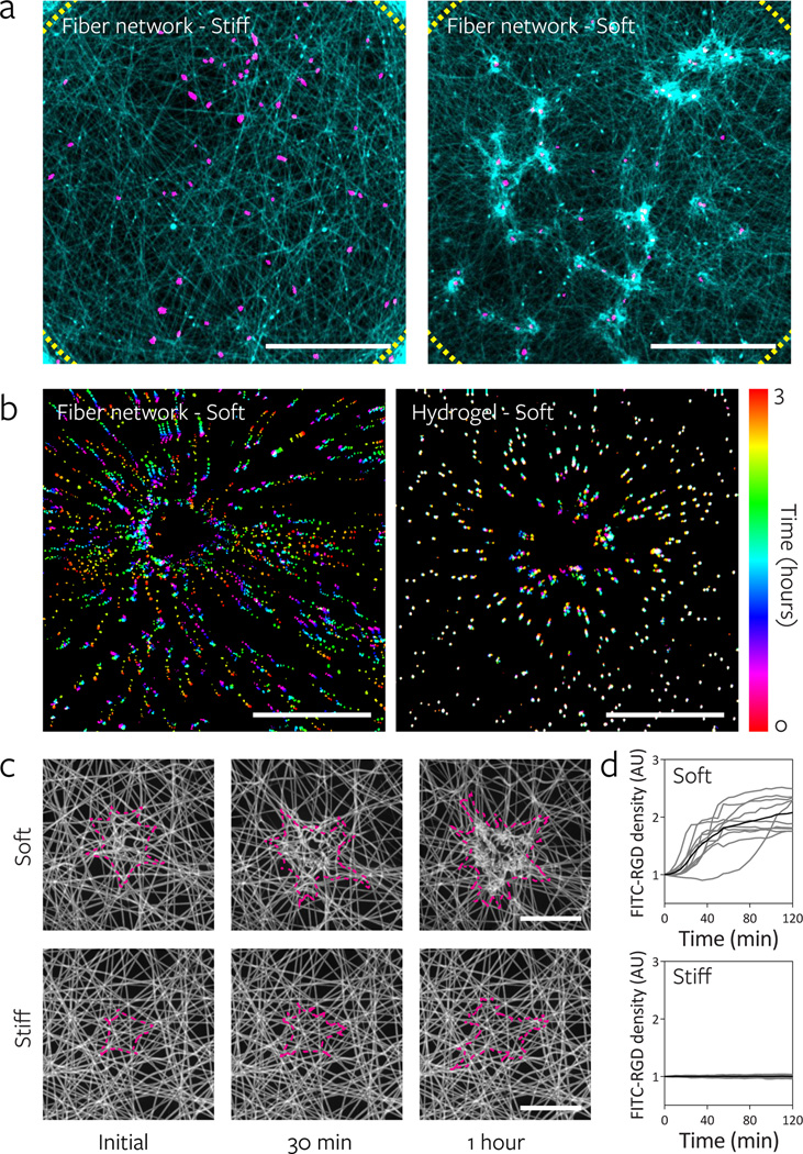
Figure 4. Lower fiber and network stiffness enables cell-mediated reorganization of the material and clustering of adhesive ligands local to the cell.
a, Reorganization of stiff and soft fiber networks 16 h after hMSC seeding. Fibers imaged by coupling with rhodamine methacrylate (cyan) and thresholded cell nuclei labeled with Hoechst 33342 (magenta). Dotted lines indicate the periphery of the suspended network (scale bars, 500 µm). b, Temporally color-coded overlays capturing the motion of beads embedded within soft fibers (fiber: 140 MPa, network: 2.8 kPa) and soft hydrogels (290 Pa) over a 3 h time course following hMSC seeding. c, Time-lapse images of FITC-RGD coupled fiber recruitment during the first two hours of hMSCs spreading on soft (top) and stiff (bottom) networks. Cell outlines shown in magenta (scale bars, 50 µm). d, Quantification of FITC-RGD fluorescence intensity in a 50 µm diameter circular region centered on the cell’s nucleus. Intensity was normalized to adjacent acellular areas; n = 10 cells.

