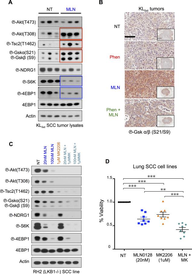
Figure 7. Co-targeting SCC with MLN0128 and MK2206.
(A) Analysis of lung tumor lysates from KLluc SCC following 8 weeks of MLN0128 treatment. Immunoblots were stained with the indicated antibodies. (B) Immunoblots from whole cell lysates from RH2 cells treated with DMSO, MLN0128 (20nM and 100nM), MK2206 (1μM), 20nM MLN0128 + 1μM MK2206 and 100nM MLN0128 + 1μM MK2206 for 24hours. Immunoblots were stained with the indicated antibodies. (C) Representative immunohistochemistry images from therapy resistant SCC tumors stained with phospho-GSKα/β (Ser21/9) antibody. Scale bars (black) = 50 μM and inset = 2mm. (D) Cell viability measured by trypan blue staining of SCC cell lines (RH2, H157, H226, H460, H520, H596, H1703, and SW900) treated with DMSO, low dose MLN0128 (20nM), MK2206 (1μM) or MLN0128 (20nM) + MK2206 (1μM) for 3 days. Statistical significance (p-values: * < 0.05; ** < 0.01; *** < 0.001;) calculated using a non-parametric one-way ANOVA (Tukey test). The data are represented as the mean ± SEM. Error bars represent the ± S.E.M.

