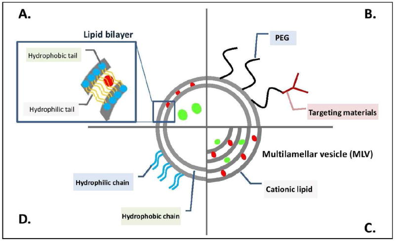
Figure 3.

Schematic diagram of various liposomal structures. (a) Illustrates the ability for liposomes to delivery both hydrophilic (green sphere) and hydrophobic (red sphere) drugs either solubilized in the core or embedded within the lipid bilayer. (b)-(d) Represent variations of the traditional liposomes to add targeting ligands or ‘stealth’ using poly(ethylene glycol). Concept of this figure is adapted from Nature Publishing Group. [55]
