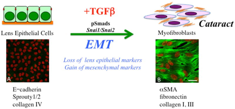
Figure 2.

TGFß induces an epithelial to mesenchymal phenotype in lens epithelial cells. In the process, cells lose many of their normal epithelial markers and characteristics as they dissociate from each other to acquire a more myofibroblastic, migratory phenotype. Some of the specific markers now expressed include alpha smooth muscle actin (α-SMA), as well as excessive levels of extracellular matrix molecules, including fibronectin and collagens type I and III. It is this cellular process that contributes to lens pathology, especially the fibrotic changes leading to polar cataracts and posterior capsular opacification. Included are representative lens epithelial explants prepared from postnatal-day-15 murine lens exposed to either no TGFß (A) or 50pg/ml TGFß2 (B) for up to 5 days, immunolabeled for α-SMA (green), with cell nuclei counterstained with propidium iodide (red). With TGFß, lens epithelial cells undergo an EMT, highlighted by α-SMA-labeled myofibroblasts. Scale bar; 20μm.
