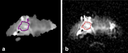
Figure 1.

ADC images for a patient with a follicular adenoma from the training set. (a) Neuroradiologist‐defined ROI of the lesion on a bitmap‐format ADC map in FuncTool. (b) The same ROI shown on the original resolution DICOM‐format ADC map in ImageJ.
