
Figure 4.
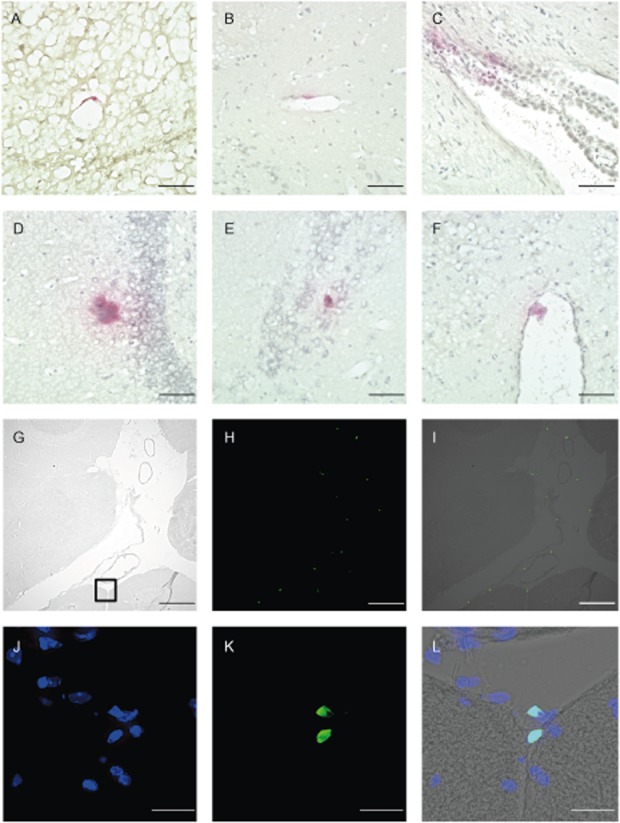
Donor cells detected in the brain of chimeric mice. Cells were stained for β-glucuronidase (GUSB) activity (A–F). (A–B) Cells at blood vessel wall, (C) Choroid plexus, (D–E) Cells in brain tissue, (F) Cells at pia mater in third ventricle, ×400. Scale bars: 50 μm (A–F). Detection of donor cells in leptomeningeal space (G–L). (G) Phase contrast microscopy, (H) green fluorescent protein (GFP) positive cells, (I) Merge, ×100. Scale bars: 200 μm (G–I). (J–L) Fluorescent microscopy. Enlarged view of the black square in (G). (J) Nuclear stained with 4′6′-diamidino-2-phenylindole dihydrochloride (DAPI), (K) GFP-positive cells at pia mater, (L) Merge. Original magnification, ×400 (J–L). Scale bars: 20μm (J-L).