
Figure 5.
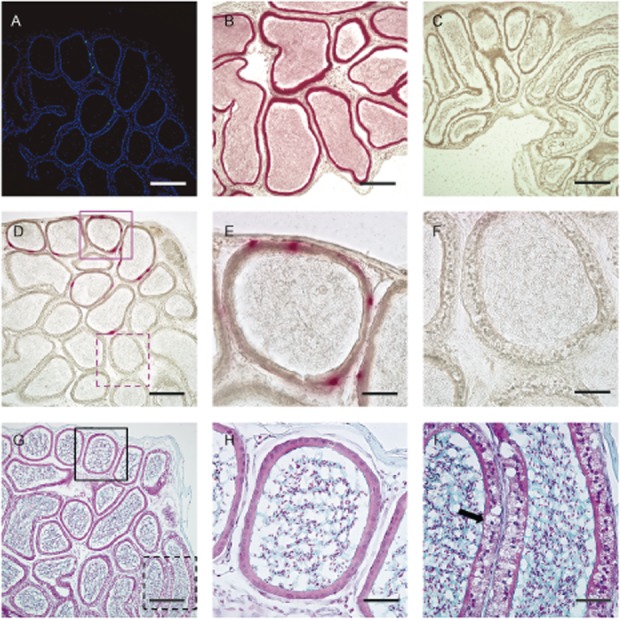
Representative staining of cross-correction by engrafted donor cells that produce β-glucuronidase (GUSB) in local areas. We analyzed frozen section slides of the epididymis sectioned close together, which were obtained from the chimeric mouse that had been rescued from infertility. (A) Fluorescent microscopy. Merge of green fluorescent protein (GFP) expressed by donor cells and nuclear, stained with 4′6′-diamidino-2-phenylindole dihydrochloride (DAPI). GFP-positive cells that had differentiated and engrafted in a limited part of the section could be seen. (B–F) Stain for GUSB activity, (B) Positive control, (C) Negative control for epididymis, ×100. Scale bars: 200 μm (A-D, G). (D) GUSB-positive cells are visible in a limited area, and the locations of some cells are the same as GFP-positive cells, shown in (A). (E–F) Enlarged views of the red-stained area (E) and the unstained area (F) from (D). (G–I) Staining with Alcian blue and counterstained with Nuclear fast red. (H–I) Enlarged views of the epididymis ducts (H, I) from (G) in a neighboring section of the red-stained area (E) and the unstained area (F). Normal appearance of epididymis ducts that contain GUSB-producing cells (E, H). Although traditional fixation and staining dissolves mucopolysaccharide in affected cells, a ballooning foamy cell appearance (arrow) can be seen in (I), ×400. Scale bars: 50μm (E–F, H–I).