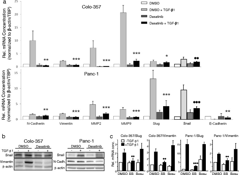
Fig. 3.

Dasatinib blocks TGF-β1-induced expression of genes involved in EMT and cell migration/invasion. a Colo-357 (upper graph) and Panc-1 cells (lower graph) cultured for 24 h in the absence or presence of TGF-β1 (5 ng/ml) and either DMSO or dasatinib (10 μM) were subjected to RNA isolation and qPCR-based determination of the indicated genes. Data are plotted relative to untreated DMSO-treated control cells (set arbitrarily at 1) and represent the mean ± SD from three independent experiments after normalisation to β-actin and TBP. Asterisks indicate a significant difference between dasatinib + TGF-β1-treated cells and DMSO + TGF-β1-treated cells (n = 3). b Colo-357 and Panc-1 cells cultured for 48 h in the absence or presence of TGF-β1 (5 ng/ml) and either DMSO or dasatinib (10 μM) were subjected to immunoblot analyses of the indicated proteins and β-actin as a loading control. One representative blot is shown for each cell line. c Colo-357 and Panc-1 cells cultured for 24 h in the absence or presence of TGF-β1 (5 ng/ml) and either DMSO, SB431542 (SB, 5 μM) bosutinib (Bosu, 10 μM) were subjected to qPCR-based determination of Slug and vimentin. Data represent the mean ± SD from three independent experiments. Asterisks indicate a significant difference between SB + TGF-β1-treated cells and DMSO + TGF-β1-treated control cells (n = 3)
