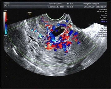
Fig. 1.

Ultrasound findings of a typical CSP. Longitudinal section of the uterus showing a 6 + 3 weeks with cardiac activity gestational sac (Case 1; crown–rump length:3.6 mm) implanted into a previous Cesarean section scar and protruding towards the urinary bladder with strong peripheral color doppler signals
