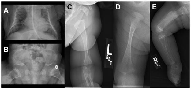
Fig. 3.
A: Small chest with nasogastric tube at 3 months of age. B: Small ilia with flat acetabular angles. C: Radioulnar synostosis of left arm. D: Right femur at 3 weeks of age with fracture and metaphyseal flaring. E: Right lower extremity at 8 months of age with healing fractures. All five radiographs show bone demineralization.

