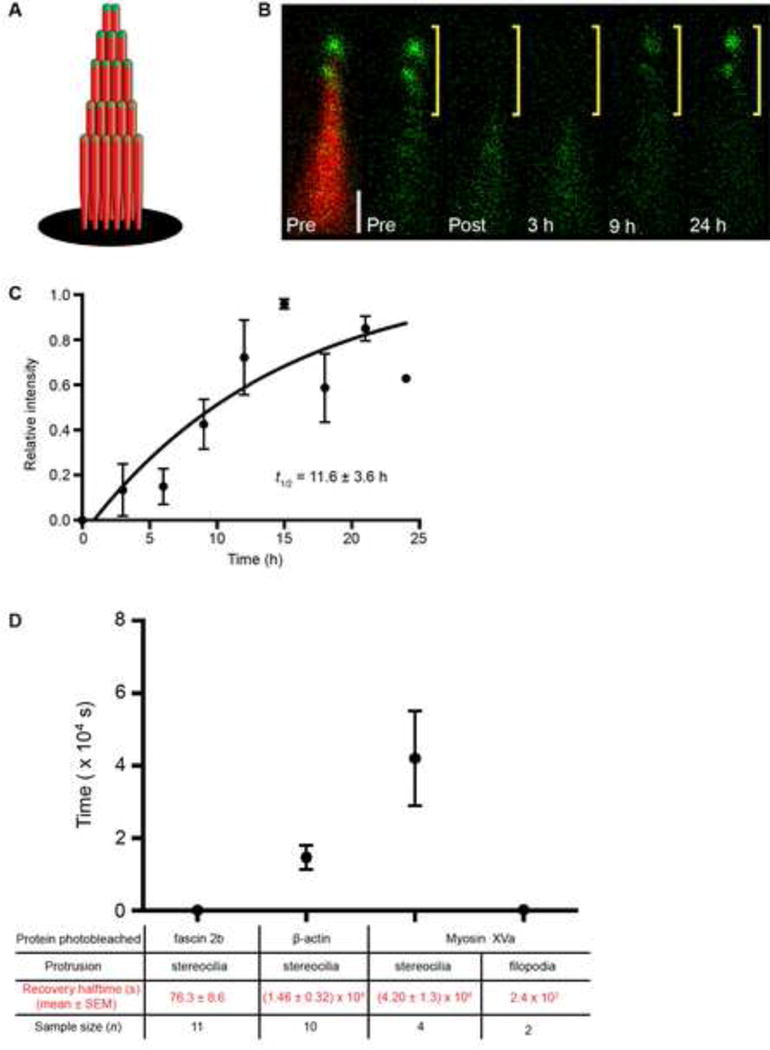
Figure 4. Robust myosin XVa exchange at the tips of mature stereocilia.

(A) Schematic of bundle containing β-actin-mCherry (red) and GFP-myosin XVa (green). (B) Image of a bundle expressing GFP-myosin XVa (green) reveals fusion protein at stereociliary tips (Pre, left and right). Bundle is counterlabeled with β-actin-mCherry (Pre, left, red) in a doubly transgenic hair cell. After photobleaching the top half of bundle (only GFP signal is shown), the GFP-myosin XVa signal recovers completely by 24 h. Scale bar is 1 μm. (C) Time course of myosin XVa recovery. Zebrafish examined at 7 dpf. (D) Summary of recovery halftimes of fusion proteins in stereocilia and filopodia. Recovery halftime of myosin XVa in filopodia is estimated (Belyantseva et al., 2005). See also Table S1.
