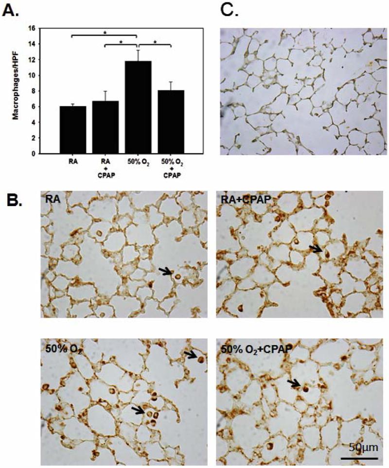
FIGURE 3.
Representative images of macrophages stained with Mac3 anti-macrophage antibodies from the four groups of pups [B] and a negative control [C] are shown together with summarized data [A]. Hyperoxic exposure increased Mac3 counts in lung parenchyma when compared to both RA groups. This increase was prevented with CPAP exposure in hyperoxic pups [*p<0.05]. The number of macrophages was counted per high power field [HPF, 20X magnification].

