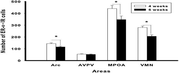
Figure 5.
Shipping female mice during pubertal development results in reduction of ER-α levels in adulthood in some brain areas. Number of ER-α IR cells (mean ± SEM) in CD-1 female mice shipped at 4 or 6 weeks old in the arcuate nucleus (Arc), anteroventral paraventricular nucleus (AVPV), medial preoptic area (MPOA) and ventromedial nucleus of the hypothalamus (VMN). (*= p < 0.05) Reprinted from (18;18) with permission of Elsevier, Copyright 2011.

