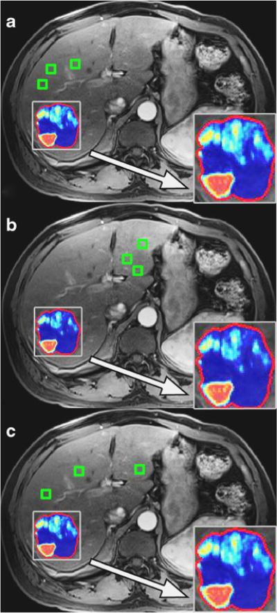
Fig. 2.

The systematic ROI placement method in the same representative patient case as in Figure 1. A) Three ROIs were subsequently placed near the lesion (i.e., ipsilateral liver lobe to the lesion). B) Three ROIs were placed away from the lesion (contralateral liver lobe to the lesion). C) Three ROIs were placed dispersed throughout the liver. Representative qEASL colour maps produced from each ROI placement method is also shown in closer detail within the zoom boxes
