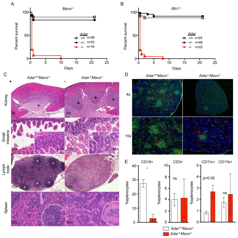
Figure 5. Postnatal mortality and severe developmental defects in Adar−/−Mavs−/− mice.
(A) Postnatal survival curves for Adar−/−Mavs−/− mice.
(B) Postnatal survival curves for Adar−/−Ifih1−/− mice.
(C) Adar−/−; Mavs−/− mice have developmental defects of the kidney (top row; papillae marked with black asterisks), small intestine (second row), lymph node (third row; follicles marked with white asterisks), and spleen (fourth row; lymphoid regions enclosed by dashed circle). Images are hematoxylin and eosin with magnification indicated.
(D) Immunofluorescence microscopy of splenic sections with anti-B220 (green), anti-CD8 and anti-CD4 (red), and DAPI (blue).
(E) Analysis of splenocytes by flow cytometry shows severe B cell deficiency in Adar−/−; Mavs−/− mice. Mice in C-D were 20 days old. Mice in E were 15 days old. Mice in F were 13 days old.

