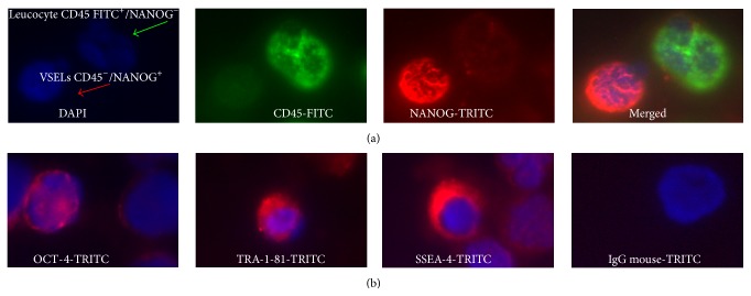
Figure 3.
Immunofluorescence analysis of PB-derived VSELs. A typical triple-staining with 4′,6-diamidino-2-phenylindole (DAPI) (blue: nuclei), fluorescein isothiocyanate- (FITC-) CD45 (green fluorescence), and tetramethylrhodamine-5-isothiocyanate- (TRITC-) SSEA-4−, TRA-1-81, OCT-4, or NANOG (red) shows (a) VSELs: small CD45− cells NANOG+ and leukocytes: greater CD45+ NANOG− cells and (b) VSELs: CD45− cells which express OCT-4 in nuclei or TRA-1-81 and SSEA-4 on the surface.

