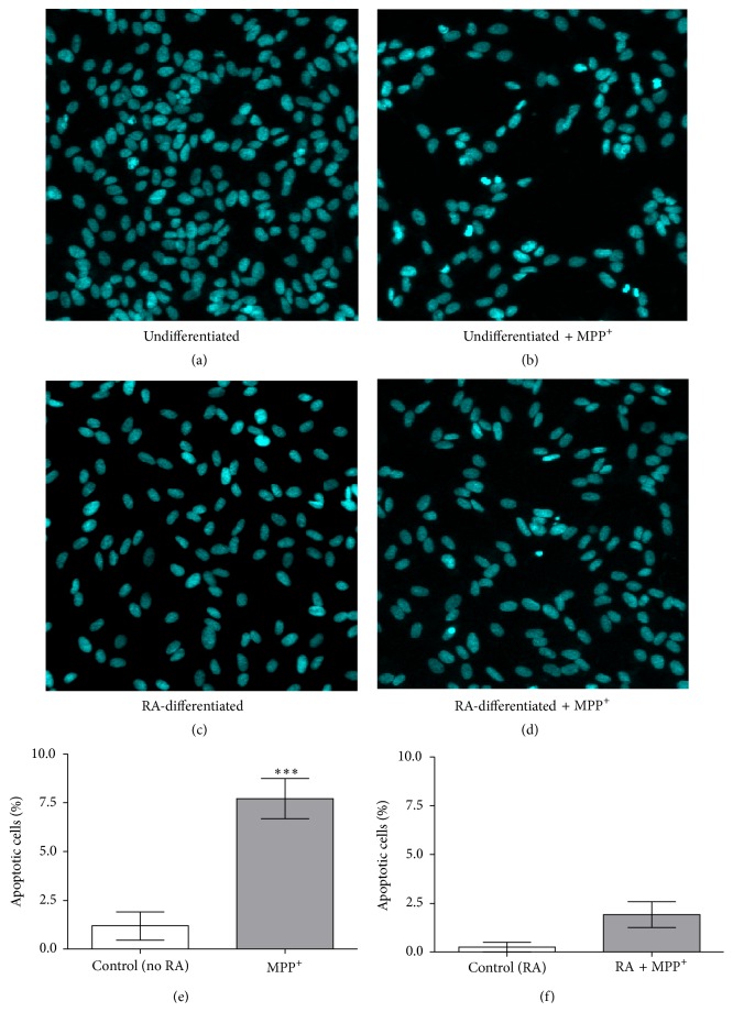
Figure 4.
Effect of MPP+ on nuclear morphology in undifferentiated and differentiated SH-SY5Y cells. Cells were undifferentiated (a and b) and were differentiated in 10-μM retinoic acid (RA) for 3 days (c and d). Undifferentiated (b) and differentiated cells (d) were exposed to 500 and 1000 μM of MPP+ for 24 hours, respectively. Apoptotic nuclear morphology was visualized by DNA staining with Hoechst 33258. Percentage of cells with apoptotic nuclei was calculated (e and f). Data are expressed as mean ± SEM (n = 3) of percentage to untreated cells. ∗∗∗ P < 0.001.

