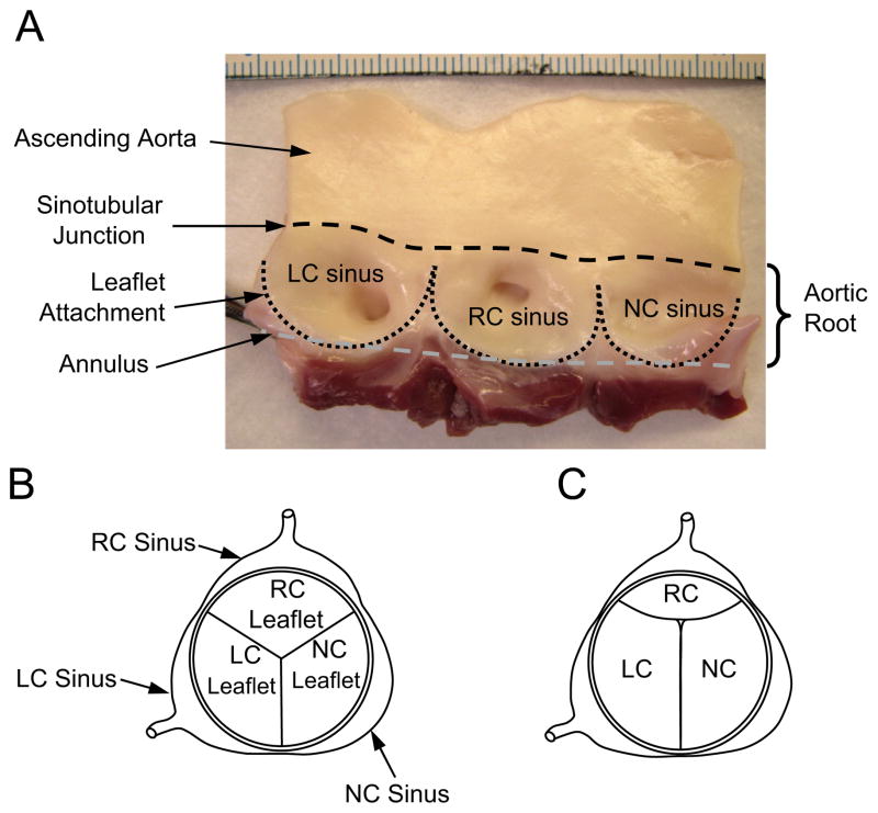
Fig. 1.
(A) Inside wall of the porcine aortic root with leaflets excised. The three sinuses, or bulges, in the aortic root are referred to as the left coronary (LC), right coronary (RC), and noncoronary (NC) sinuses, the former two serving as the origins of their respective coronary arteries. The juncture of the sinuses with the ascending aorta is referred to as the sinotubular junction (black dashed line). The leaflet attachment serves as the hemodynamic boundary between the left ventricle and the aorta (black dotted line). The aortic annulus is used here to describe the plane that passes through the nadirs of the leaflet attachments (gray dashed line). Scale (top of photo) is in millimeters. (B) Top view of aortic valve showing a closed valve in which the three leaflets and sinuses are approximately equal in size. (C) Aortic valve in which the right coronary (RC) leaflet and sinus are significantly smaller than the left coronary (LC) and noncoronary (NC) leaflets and sinuses.

