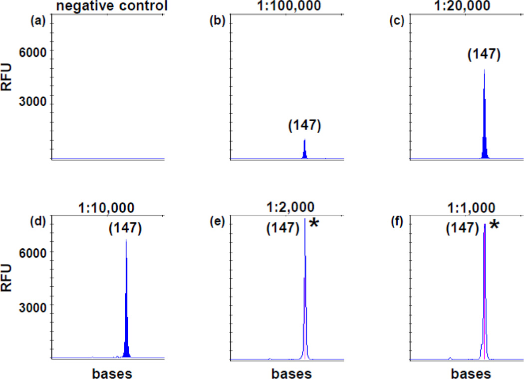
Fig 2. Analytic sensitivity of TD-PCR for primer pair TD3.
DNA from a diagnostic FLT3/ITD AML of nearly 100% leukemia cells was serially diluted with normal DNA: 1 in 1,000 (10−3 in f), 1 in 2,000 (5×10⁻⁴ in e), 1 in 10,000 (10−4 in d), 1 in 20,000 (5×10⁻⁵ in c) and 1 in 100,000 (10−5 in b). (b) is equivalent to approximately 0.4 mutant cell genomes in 250 µg DNA. (a) is normal DNA. PCR products from (e) and (f) were diluted 10-fold before electrophoresis. “*” indicates off-scale peaks. Sizes in bases are in parentheses. RFU: relative fluorescence unit.

