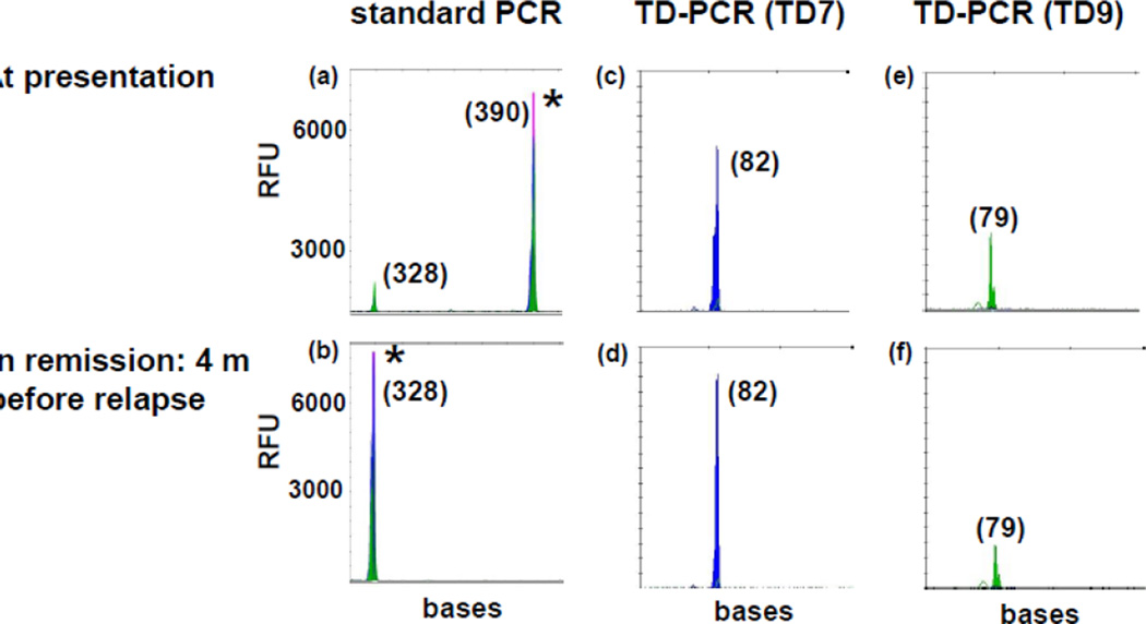
Fig 3. TD-PCR detected MRD at 4 months before relapse.
Standard PCR showed an ITD mutant migrating at 390 bases at both presentation (a) and relapse (not shown), but not at 4 months before relapse (b). TD-PCR using primer pair TD7 (c and d) detected MRD in remission (d). The ITD mutant was detected in 1 of 4 replicates of 250 ng DNA per reaction and in 1 of 20 replicates of 50 ng DNA per reaction (data not shown), indicating MRD at a level of approximately one in 160,000 cells. Duplication-specific amplification was also confirmed by another primer pair TD9 (e and f). TD-PCR products at presentation (c and e) were diluted 100 fold before electrophoresis. ‘*” indicates off-scale peaks. Sizes in bases are in parentheses. RFU: relative fluorescence unit.

