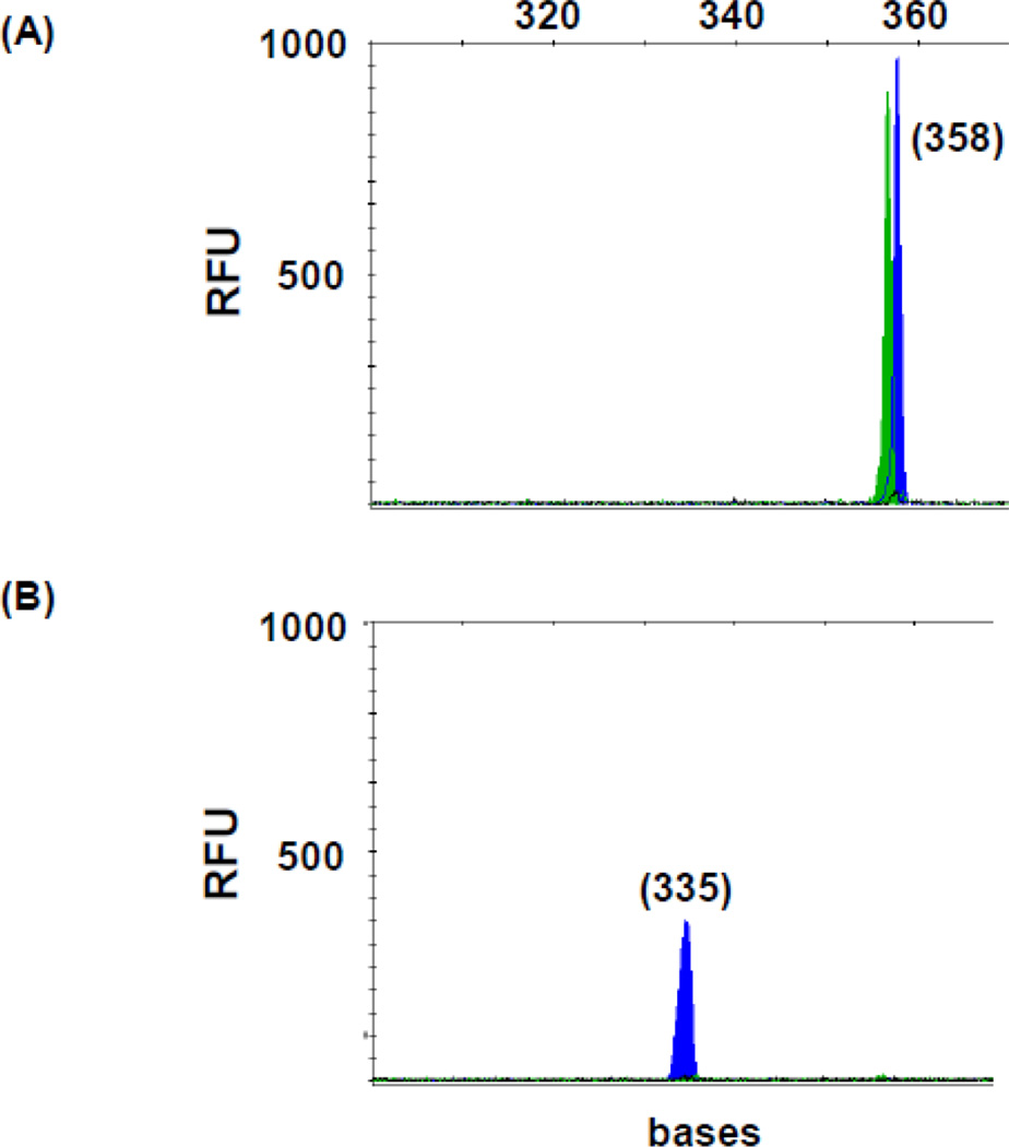
Fig 4.
FLT3/ITD mutation in normal marrow. TD7 TD-PCR detected an ITD migrating at 358 bases (A). Duplication-specific amplification was confirmed by ScaI restriction enzyme digestion with a peak migrating at 335 bases (B) and by Sanger sequencing. Sizes in bases are in parentheses. RFU: relative fluorescence unit.

