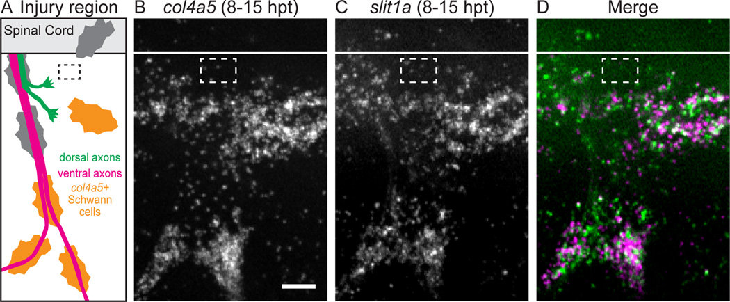
Figure 6. Slit1a is upregulated with collagen4a5 after nerve transection.
(A) Schematic showing approximate region imaged for in situ hybridization (black dashed box, transection site) (B–D) col4a5 mRNA (B) and slit1a mRNA (C) are co-expressed (D) ventral to the spinal after nerve transection (n = 6 larvae, 27/30 nerves). White line, dorsal aspect of spinal cord; white dashed box, approximate transection site; scale bar = 10 µm.

