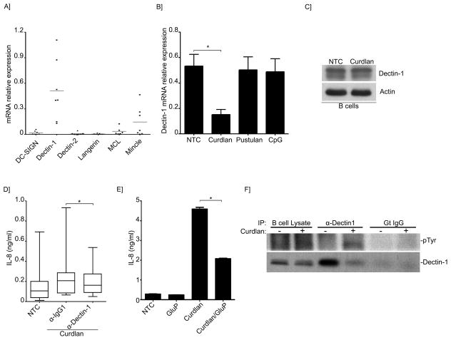
Figure 3. Dectin-1 participates in IL-8 secretion induced by β-glucans.
(A) Quantitative real-time PCR for the indicated C-lectin receptors mRNAs in B-lymphocytes. Expression was normalized to GAPDH. Shown are the data from 7 different B-cell preparations. (B) Quantitative real-time PCR for Dectin-1 mRNAs in B-lymphocytes of NTC, or after stimulation with Curdlan, pustulan or CpG. Expression was normalized to GAPDH. Shown are the data from 3 different B-cell preparations. *p< 0.05 (C) Immunoblot analysis of Dectin-1 in unstimulated or Curdlan-stimulated B-lymphocytes. (D)IL-8 was measured by ELISA in the supernatant of NTC and after Curdlan stimulation. Cells were pretreated with Dectin-1 neutralizing antibodies or isotype control as indicated. Data were analyzed by the non-parametric Wilcoxon matched-pairs signed rank test to compare the medians of 5 different B-cell preparations. (E) Secretion of IL-8 into the cell supernatant was measured by ELISA of NTC or Curdlan-stimulated cells. Cells were pretreated with glucan phosphate as indicated. Shown is a representative run of 3 independent experiments performed with 3 different B-cell preparations. *p< 0.01. (F) Endogenous Dectin-1 was immunoprecipitated from unstimulated and Curdlan-stimulated cells phospho-tyrosine antibody was used to detect Dectin-1 phosphorylation in the immunoprecipitates. Total Dectin-1 was used as a control.

