
Figure 6. Blocking of fusion with antibody.
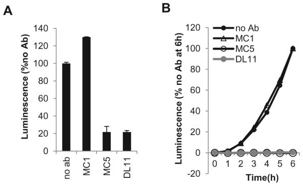
(A). Firefly Luciferase Assay. One population of CHO-K1 cells was transfected with the gD, gH, gL, gB and T7 polymerase plasmids. The other population was transfected with receptor and luciferase plasmids. 6 hours post-transfection the two populations were co-cultivated and 100μg/ml of antibody (MC1, MC5 or DL11) was added. 18 hours post co-cultivation, cells were lysed and luminescence measured. Values were normalized to no antibody control. (B). Split Luciferase Assay. B78 cells were transfected with gB, gH, gL, and one of the DSP plasmids. C10 cells were transfected with the other DSP plasmid. An hour prior to co-cultivation, 30μg/ml soluble gD was pre-incubated with 30μg/ml of either MC1, MC5 or DL11. The gD-Ab mixture was then added at the co-cultivation step. Fusion was monitored over 6 hours. Data were normalized to no antibody control.