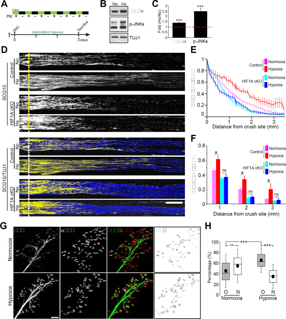
Figure 8. Acute intermittent hypoxia (AIH) enhances axon regeneration in vivo.
(A) Experimental scheme for in vivo AIH: 6 10min hypoxic episodes (8% oxygen) with equivalent 10min normoxic intervals. Green bar indicates 10 min hypoxic condition and black bar indicates 10 min normoxic condition (AIH total duration=120 min). Control mice followed the same 120 min regime under normoxia. SNC: sciatic nerve crush. (B) Western blot analysis of mouse L4 and L5 DRGs treated for 120 min of normoxia (No) or hypoxia (Hx) using the AIH protocol. (C) Quantifications of protein level of HIF-1α and p-JNKs (n=3 for each condition; ***p<0.001, **p<0.01 by t-test; mean±SEM). (D) Representative longitudinal sections of sciatic nerves from control or HIF1AcKO mice, dissected 3 days after injury with or without daily AIH and immunostained for SCG10 and βIII tubulin. Scale bar, 500µm. (E and F) In vivo regeneration index calculated from (D) (n=7 and 10 for normoxia and hypoxia from control mice, n=10 and 8 for normoxia and hypoxia from HIF1AcKO mice; *p<0.05 by one-way ANOVA with Tukey test; mean±SEM; ns, not significant). (G) Motor axons reinnervation assays. EHL muscles of thy1-YFP-16 mice dissected 12 days after sciatic nerve injury with or without daily AIH were stained with Alexa Fluor 647-conjugated α-Bungarotoxin (αBTX). Scale bar, 100µm. (H) Quantification of percentage of axon-non-occupied (N) and axon-re-occupied NMJ end plates (O) (n=15 for each condition; box, 25 to 75%; whisker, standard deviation; closed circle, mean; ***p<0.001 by one-way ANOVA with Tukey test; ns, not significant).

