Figure 8. Proposed model of allosteric regulation of opioid receptors by the α 2A-adrenoceptor.
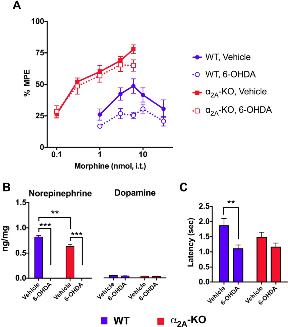
A) Schematic showing dose-response curves obtained for spinal morphine under different experimental conditions. In WT mice, morphine produces a dose-dependent antinociceptive effect (blue solid line), which is reduced in mice treated with 6-OHDA (blue dashed line) and potentiated by the addition of clonidine (purple solid line). In α2A-KO mice, morphine is more potent than in WT mice (red solid line) and neither the 6-OHDA treatment (red dashed line) nor the addition of clonidine (purple dashed line) shifted the morphine dose-response curve. B) In the proposed model, in WT mice, opioid receptors (OPr, green) are inhibited by the α2A-AR (red) in its ligand-free inactive state, which results in decreased opioid receptor antinociceptive output. When an agonist like clonidine (Clon) or norepinephrine (NE) activates α2A-AR, this inhibitory action on opioid receptors is removed, resulting in the synergism between the two agonistoccupied receptors. Under normal conditions, tonic NE release from descending noradrenergic fibers maintains an equilibrium state between ligand-free and agonist-occupied α2A-AR. When NE is depleted, the equilibrium is shifted towards the inactive and inhibitory α2A-AR, and when exogenous clonidine is administered, the equilibrium is shifted towards an active α2A-AR state, which disinhibits the opioid system. In α2A-KO mice, opioid receptors are not subjected to inhibition by the α2A-AR regardless of the presence or absence of α2A-AR ligands and are thus always fully activated.

