Figure 4.
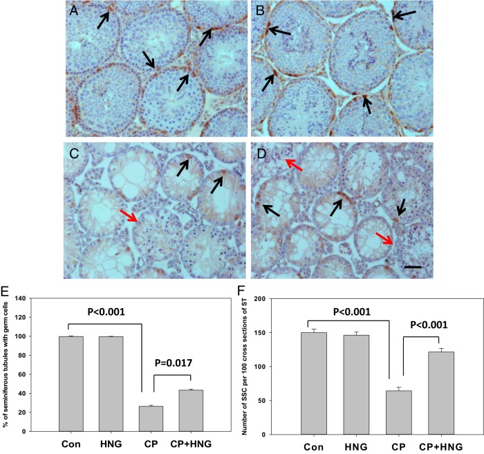
Representative immunohistochemistry micrographs of mouse testis sections from control (n = 5) (A), HNG daily sc injection (n = 5) (B), 6 repeated dose of CP treatment (n = 10) (C), and CP+HNG-treated mice (n = 10) (D); scale bar, 0.05 mm. The cross-sections of seminiferous tubules containing elongated spermatids (red arrows) were noted both in CP (C) and CP+HNG (D)-treated mice. The FoxO1-positive SSCs (dark brown in color, black arrows) were observed in testes from all groups. The bar graph (E) shows the percentage of seminiferous tubules containing germ cells in control, HNG-treated alone, repeated CP alone, and combined CP with HNG-treated (CP+HNG, n = 10) mice. The bar graph (F) shows the number of SSCs (FoxO1-positive spermatogonia) per 100 cross-sections of seminiferous tubules from control (Con), HNG-treated alone, repeated CP alone, and combined CP with HNG-treated (CP+HNG) mice. Values are mean ± SEM.

