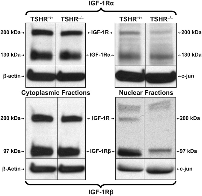
Figure 4.
Western blot analysis of IGF-1R content in cytoplasm and nucleus of TSHR+/+ and TSHR−/− fibroblasts. Fibroblasts were subjected to subcellular fractionation and proteins extracted, as described in Materials and Methods. Proteins were loaded on 4%–20% triglycine gels and subjected to SDS-PAGE. Separated proteins were then probed by Western blotting with anti-IGF-1Rα and anti-IGF-1Rβ Abs.

