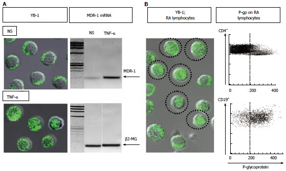
Figure 1.

Up-regulation of nuclear translocation of Y-box-binding protein-1, transcription of multidrug resistance 1 in lymphocytes, and P-glycoprotein expression on lymphocytes. A: Left: Immunostaining and confocal microscopy analysis of Y-box-binding protein-1 (YB-1) in 1 × 10⁵ of peripheral blood mononuclear cells (PBMCs). YB-1 was expressed in the cytoplasm of all non-stimulated PBMCs (NS). In contrast, nuclear translocation of YB-1 was induced in 30% or more of PBMCs incubated with 10 ng/mL of tumor necrosis factor-α (TNF-α). Immunostaining for YB-1 using a specific antibody (Ab) against YB-1[19] with FITC-conjugated anti-rabbit IgG Ab (BD Biosciences Pharmingen). Confocal analysis of YB-1 using a LSM 5 Pascal invert Laser Scan Microscope (Carl Zeiss Microscope Systems, Germany). Magnification, × 600; Right: Multidrug resistance-1 (MDR-1) mRNA expression was examined by RT-PCR using total RNA extracted from PBMCs incubated with 10 ng/mL of TNF-α or no stimulation (NS). The primer sequences were as follows: human β2-microglobulin forward 5’-ACCCCCACTGAAAAAGATGA-3’, reverse 5’-ATCTTCAAACCTCCATGATG-3’; human MDR-1 forward 5’-CCCATCATTGCAATAGCAGG-3’, reverse 5’-GTTCAAACTTCTGCTCCTGA-3’. Amplified products were electrophoresed with Marker 4 (Nippon Gene, Tokyo) on 3% agarose gels; B: Spontaneous nuclear translocation of YB-1 and P-glycoprotein (P-gp) expression on lymphocytes from a typical patient with active rheumatoid arthritis (RA). Left: Immunostaining and confocal microscopy analysis of YB-1 in 1 × 10⁵ of PBMCs. YB-1 was expressed in the nuclei of a proportion of unstimulated PBMCs (encircled cells). Magnification, × 600; Right: P-gp expression on CD4+ and CD19+ peripheral blood lymphocytes. The dotted line represents the gate set to discriminate negative from positive stained cells as determined by control FITC-conjugated anti-mouse IgG Ab. The specific antibodies for staining and flow cytometric analysis were as follows: staining for P-gp using MRK16 (a specific monoclonal Ab against P-gp; Kyowa Medex, Tokyo) with FITC-conjugated goat anti-mouse IgG Ab (BD Biosciences Pharmingen), cy-chrome-conjugated CD4 monoclonal Ab, cy-chrome-conjugated CD19 monoclonal Ab (BD Biosciences Pharmingen).
