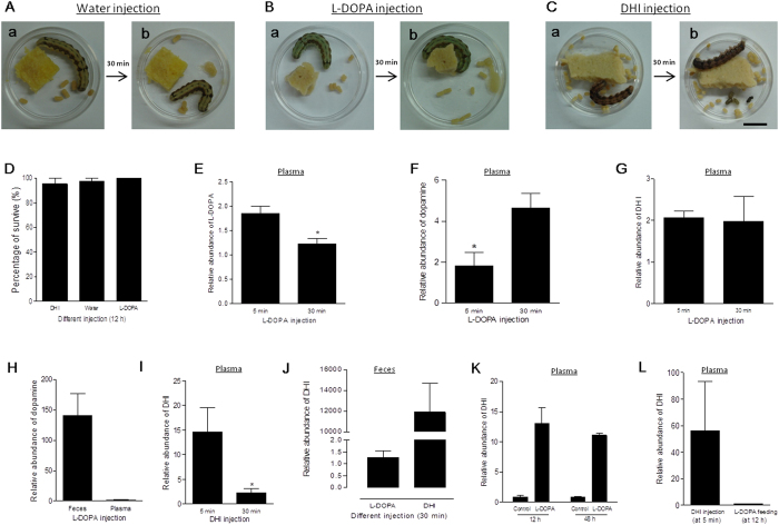
Figure 9. Hemolymph DHI was transferred into the hindgut for excretion without causing cytotoxicity in vivo.
Identical volumes of sterile water (A), l-DOPA (B), and DHI (C) were injected into H. armigera larvae (V-1) respectively. The excreted feces were compared before and 30 min after injection. (D) The survival rates of H. armigera larvae did not differ among the treatments. (E–H) l-DOPA in hemolymph introduced through injection was metabolized into dopamine but not DHI through DDC. H. armigera larvae were injected with l-DOPA. At 5 and 30 min post injection, plasma was obtained to determine plasma l-DOPA (E), dopamine (F), and DHI (G) levels. (H) At 30 min, the dopamine levels in feces and plasma were also compared. (I–K) DHI is not cytoxicity in vivo. (I–J) DHI was excreted from hemolymph into feces. H. armigera was injected with DHI as shown in (C). At 5 and 30 min post injection, plasma was obtained to determine DHI (I). (J) The relative amounts of DHI in the feces from larvae that received l-DOPA and DHI injections were compared. (K) The DHI levels in larval plasma were determined after being fed a normal diet (control) or a diet supplemented with l-DOPA at 12 and 48 h, respectively. (L) Plasma DHI levels were compared between H. armigera larvae that underwent DHI injection at 5 min or l-DOPA feeding for 12 h. Each column represents the mean of three independent measurements ± S.E.M. *P < 0.05. Bar: 1 cm.

