
Figure 1. Crystal structure of Ube2K.
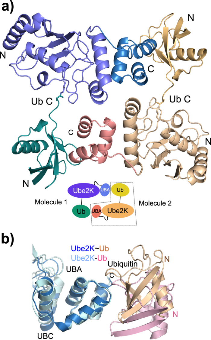
(a) Cartoon representation of the crystal structure of the Ube2K~Ub conjugate and one of its symmetry mates highlighting the interaction between the molecules. Inset: schematic of the structure indicating the two molecules. (b) Overlay of the UBA domain of the Ube2K conjugate (blue and beige) and the Ube2K:Ub complex (pale blue and pink; PDB ID: 3K9P) showing the shift in the position of ubiquitin.