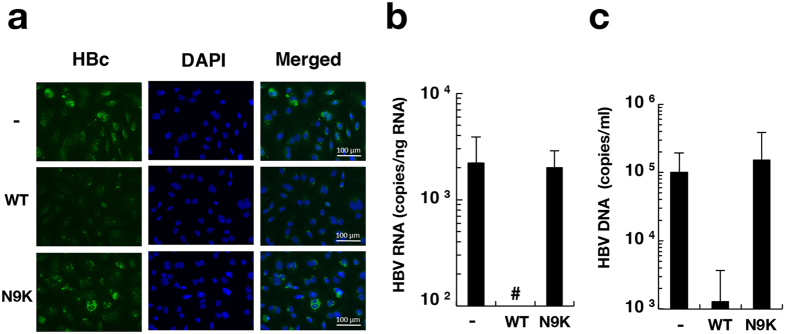
Figure 6. Inhibitory effect of preS1 lipopeptide on HBV infectivity.
HepG2/NTCPA3 cells were infected with HBV at 1,000 GEq/cell under non-adherent conditions in the presence or absence of myr-47WT (WT) or myr-47N9K (N9K). (a) The infected cells were fixed at 12 dpi. The fixed cells were stained with an antibody to HBc and counterstained with DAPI. (b) The infected cells were harvested at 6 dpi. The amount of intracellular HBV RNA was quantified by real-time qRT-PCR. (c) The culture supernatants were harvested at 12 dpi. The amount of supernatant HBV DNA was quantified by real-time qPCR. Data were calculated from 4 to 6 independent experiments and are expressed as means ± SD. Number sign (#) indicates “a value was not detected”.

