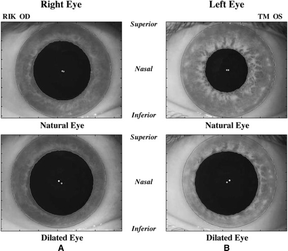
Figure 4.

Changes in pupil center location and iris shape with pupil dilation. These images illustrate the change in pupil center location and iris shape from a natural undilated state to a dilated state in (A) one patient’s right eye and (B) a different patient’s left eye. Superior, nasal, and inferior directions are noted on the figure. White and gray filled circles denote limbus and pupil centers, respectively. Irises tended to thin more in the inferonasal direction than in the superotemporal direction. Pupil centers tended to shift in the inferonasal direction with dilation. (Reprinted from J Cataract Refract Surg, Vol 32, Issue 1, Porter J, Yoon G, Lozano D, Wolfing J, Tumbar R, Macrae S, Cox IG, Williams DR, Aberrations induced in wavefront-guided laser refractive surgery due to shifts between natural and dilated pupil center locations, Pages 21–32, Copyright © 2006. published with permission from Elsevier.).
