Fig. 1.

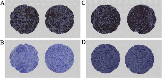
Photographs of TGF-β and PTHrP expression in breast cancer tissues by immunohistochemical staining. a.b Representative images of TGF-β positive (a) or TGF-β negative (b) cases with immunostaining (magnification × 200); c.d Representative images of PTHrP positive (c) or PTHrP negative (d) cases with immunostaining (magnification × 200)
