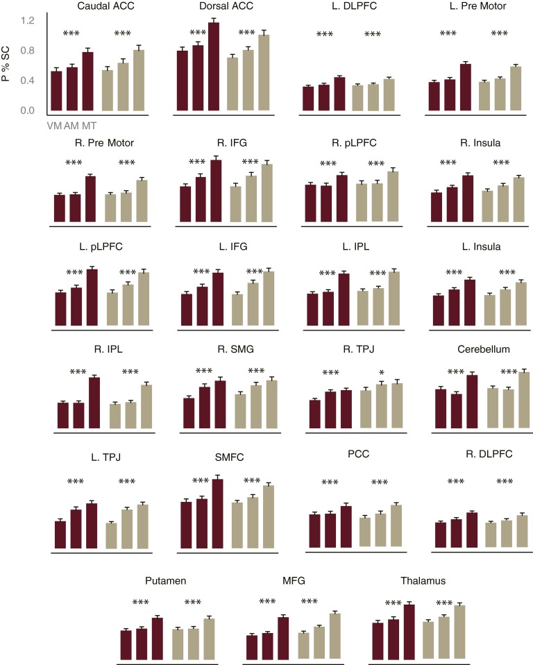
Fig. S3.
Peak percent signal change for multitask and single-task conditions. We first identified FP-SC brain regions that showed increased BOLD activity for the shape single-task and for the sound single-task (conjunction analysis), as would be expected of a task-general brain area. These ROIs were defined across all participants (n = 100) in the pretraining session. This analysis implicated a wide range of frontal, parietal, and subcortical regions. To examine which of these task- general ROIs were involved in multitasking, we identified the areas that showed a larger peak percent signal change for the multitask condition relative to the mean of the two single-task conditions, using extracted time course data. To ensure that evidence for any region’s multitasking involvement replicated across both groups of participants (internal replication), this analysis was performed separately for both the training (n = 50) and control (n = 50) groups at the pretraining session. All of the task-general ROIs showed evidence for involvement in multitasking, suggesting that neurons coding various aspects of the concurrent task performance are more widespread throughout the brain than has been previously hypothesized (3, 20). Error bars reflect SEM. ACC, anterior cingulate cortex; DLPFC, dorsal lateral prefrontal cortex; IFG, inferior frontal gyrus; IPL, inferior parietal lobule; L, left; MFG, medial frontal gyrus; P % SC, peak percent signal change; PCC, posterior cingulate cortex; pLPFC, posterior lateral prefrontal cortex; R, right; SMFC, superior medial prefrontal cortex; SMG, superior medial gyrus; TPJ, temporal parietal junction. Talaraich coordinates are presented in Table S1. *P < 0.02; ***P < 0.001.

