Fig. 3.
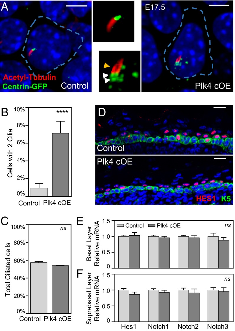
Centrosome amplification induces morphological defects in cilia in a small fraction of cells, with no global effect on Notch signaling. (A) Immunofluorescence images with enlarged panels of E17.5 whole-mount epidermis depicting cells with extra centrosomes that have a second cilium emanating from a nearby centriole. White arrowheads point to extra centrosomes; the yellow arrowhead points to second cilium. Blue dashed lines mark cell boundaries based on E-cadherin staining. DAPI is in blue. (Scale bar: 5 μm.) (B) Quantitation of the number of cilia per basal cell; 320 control and 365 Plk4 cOE cells were counted from at least two embryos. Error bars indicate SEM.) (C) Quantitation of the total number of ciliated cells in the basal layer; 161 control and 180 Plk4 cOE cells were counted from at least two embryos. Error bars indicate SEM. (D) Immunofluorescence images of HES1 within the E17.5 epidermis demonstrates unperturbed Notch pathway activation. DAPI is in blue. (Scale bar: 20 μm.) (E and F) Quantification of Notch by qRT-PCR of purified basal epidermal cells (E) and suprabasal cells (F) from 12 control and 4 Plk4 cOE embryos. Error bars represent SEM.

