Fig. 4.
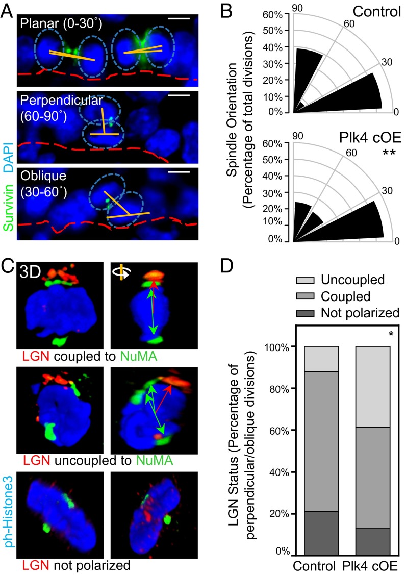
Centrosome amplification induces defects in spindle orientation by uncoupling the spindle from cortical cues. (A and B) Immunofluorescence images (A) and quantification (B) of IFE basal cells undergoing planar, perpendicular, and oblique divisions, as determined by the angle of spindle orientation (90 control and 79 Plk4 cOE divisions from three embryos each). The proportion of perpendicularly oriented spindles is skewed toward oblique orientations in Plk4 cOE tissue. Cells were stained for β4-integrin to mark the basement membrane, and with survivin to mark the linkage between two dividing cells in either anaphase or telophase. Spindle orientation was measured by the relative angle (orange lines) between the two centers of mass of DNA (blue ovals) and the basement membrane (red dashed lines). Statistical significance was established with Fisher’s exact t test. (Scale bar: 5 μm.) (C and D) Immunofluorescence images with 3D confocal reconstruction (C) and quantification (D) of LGN crescents and relative NuMA localization (69 control and 67 Plk4 cOE mitotic cells from three embryos each.) In wt skin, asymmetric cell divisions, accompanied by NOTCH/HES1 suprabasal signaling, occur when the two are coupled. In Plk4 cKO skin, there is a greater fraction of cells in which the two are uncoupled. Green double-headed arrows represent the orientation of spindle poles, and red arrows represent the corresponding alignment with the cortical LGN crescent. Statistical significance was established with Fisher’s exact t test.

