Fig. 5.
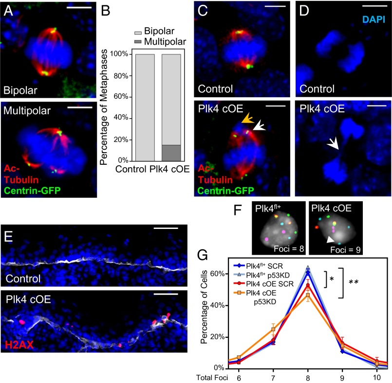
Centrosome amplification induces mitotic errors during cell division in the IFE, resulting in DNA damage and aneuploidy. (A, C, and D) Immunofluorescence images of mitotic cells in E17.5 whole-mounted epidermis. Images highlight multipolar mitoses in cells with extra centrosomes (A), misaligned chromosomes (yellow arrow) resulting from the formation of bipolar spindles when extra centrosomes cluster together (white arrow) (C), and chromatin bridges (white arrow) of sheared chromosomes (D). DAPI is in blue. (Scale bar: 5 μm.) (B) Quantification of mitotic cells at the metaphase stage in either bipolar or multipolar configuration; 32 control and 33 Plk4 cOE mitotic cells, from two embryos each. (E) Immunofluorescence images of epidermis stained for γ-H2AX as a measure of DNA damage. DAPI is in blue; β4-integrin is in white. (Scale bar: 40 μm.) (F and G) Representative images (F) and quantification (G) of FISH analysis using four chromosomal probes to gauge aneuploidy, measured by the presence of more than two or less than two foci per probe (white arrowhead); 100+ cells scored from each of three embryos per genotype/shRNA. Plk4 cOE cells have an increased frequency of aneuploid cells, which is further exacerbated when p53 levels are reduced. Error bars represent SEM.

