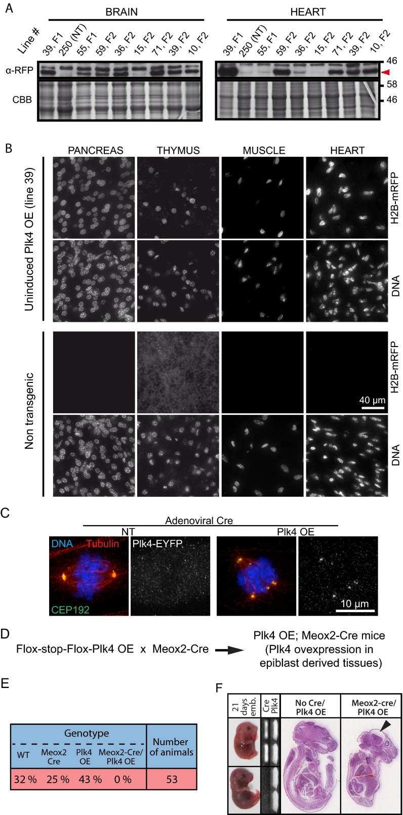
Fig. S1.
Chronic Plk4 overexpression beginning early in embryogenesis is lethal. (A) Immunoblots of brain and heart tissue lysates obtained from different inducible Plk4 mouse lines. Numbers indicate the founder line. Note that the Right panel presenting heart tissue lysates also appears in Fig. 1B. NT, nontransgenic. Red arrowhead indicates the mRFP signal. (B) Immunofluorescence images of tissues from nontransgenic control or the Plk4 OE mouse line (line 39). H2B-mRFP–positive nuclei are present in all tissues anlayzed. (Scale bar: 40 μm.) (C) Immunofluorescence images showing mitotic spindles in nontransgenic and Plk4 OE MEFs at 2 d after Ad-Cre transduction. (Scale bar: 10 μm.) (D) Outline of the breeding scheme used to obtain Meox2-Cre;Plk4 OE animals. (E) The Plk4 transgenic line 39 was crossed to mice expressing Cre under the control of the Meox2 promoter. The table shows the proportion of animals obtained with the indicated genotypes. No Meox2-Cre;Plk4 OE animals were obtained, indicating that early and ubiquitous Plk4 overexpression leads to embryonic lethality. (F) Meox2-Cre;Plk4 OE embryos where collected at 21 d postfertilization. (Left) Images documenting delayed development in Meox2-Cre;Plk4 OE embryos. (Middle) PCR genotyping of the embryos. (Right) H&E staining of sections of control (no Meox2-Cre; Plk4 OE) and Meox2-Cre;Plk4 OE embryos. The black arrowhead points to microcephaly in the Meox2-Cre;Plk4 OE embryo.

