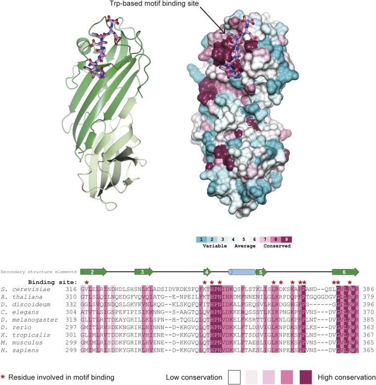
Fig. S3.
Conservation of the tryptophan-based motif-binding site in δ-COP MHD. The Consurf server (consurf.tau.ac.il/) was used to create a surface representation of evolutionary conservation of residues in the δ-COP MHD (also shown in Fig. 1D), based on an alignment from yeast to humans using ClustalO (shown here). The region that forms the tryptophan-based motif-binding site in this alignment is shown here. Residues directly involved in the binding site are highlighted above the alignment in red. The tryptophan-based motif-binding site is the outstanding feature of the conservation surface representation, indicating its importance. The Dsl1p WxW peptide is shown. Note the conservation of the length of the loop between strands 5 and 6.

