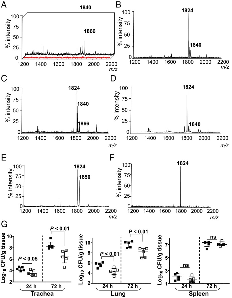
Fig. 1.
Analysis of K. pneumoniae lipid A in vivo. Negative ion MALDI-TOF mass spectrometry spectra from: (A) BALF obtained from mock-infected animals (red spectrum) or Kp52145-infected mice (black spectrum); (B) Kp52145 grown in LB; (C) from bacteria recovered after plating the infected lung homogenate in LB agar plates for 24 h at 37 °C; (D) bacteria recovered after second passage of bacteria isolated from infected lung homogenate; (E) BALF obtained from lpxO-infected mice; (F) lpxO mutant grown in LB; and (G) bacterial loads in the tissues of infected mice (n = 5 per time point and strain). Kp52145, black symbols; 52145-ΔlpxO, white symbols. Results are reported as log colony-forming units per gram of tissue (log cfu/g). ns, not significant (P > 0.05; one-tailed t test). In A and E, results are representative of five indepedent extractions; in B–D and F, results are representative of extractions from 10 infected animals.

