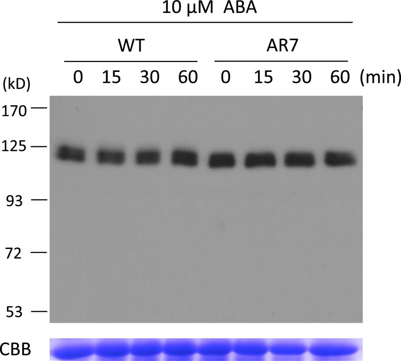
Fig. S1.
(Upper) Immunoblot analysis of the WT line and AR7 for detection of ARK. Protonemata were incubated with 10 µM ABA for the indicated times and were homogenized in a buffer containing 50 mM Tris⋅Cl (pH 8.0), 150 mM NaCl, and 1% Triton X-100 with a 1/100 volume of proteinase inhibitor mixture (P9599; Sigma,) on ice. After centrifugation at 14,000 × g for 10 min at 4 °C, the supernatants were recovered and used for electrophoresis. The proteins (30 µg) were electrophoresed on a 6% (wt/vol) SDS-polyacrylamide gel, blotted onto a PVDF membrane, and reacted with affinity-purified anti-ARK antibody. (Lower) Coomassie Brilliant Blue staining of the large subunit of the ribulose 1,5-bisphosphate carboxylase/oxygenase.

