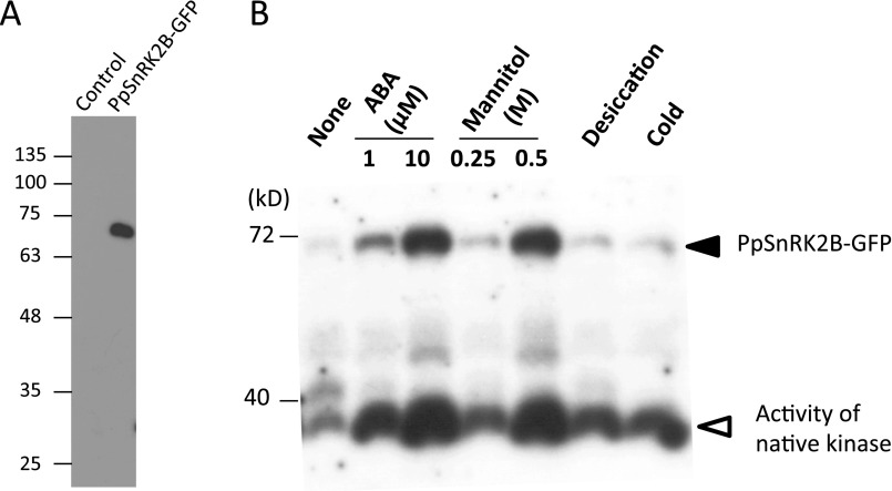
Fig. S4.
Analysis of transgenic P. patens expressing PpSnRK2B-GFP. (A) Immunoblot analysis of control and transgenic P. patens expressing PpSnRK2B-GFP using the anti-GFP antibody. (B) In-gel kinase assay of protonemata of transgenic P. patens treated with ABA (1 or 10 µM, 30 min), mannitol (0.25 or 0.5 M, 30 min), desiccation (30 min), or cold (1 h). Electrophoresis was carried out using a polyacrylamide gel containing histone IIIS as a substrate. After denaturation and renaturation treatment, the proteins were reacted with γ-32P ATP. After washing, the gel was dried and exposed to X-ray film. Note that the film is overexposed for native kinase signals for better visualization of PpSnRK2B-GFP signals.

