. 2014 Sep 12;5(12):1779–1788. doi: 10.1039/c4md00269e

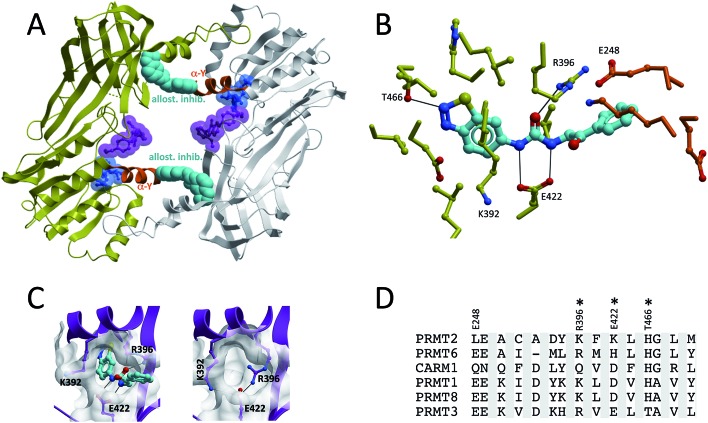
Fig. 7. Allosteric inhibition of PRMT3. (A and B) An allosteric PRMT3 inhibitor binds in a cavity of the β-barrel, at the base of the dimerization arm, and is buttressed against the α-helix (orange) of the other PRMT3 subunit. The cofactor-binding pocket is highlighted by a cofactor molecule (blue), and the substrate binding pocket by a substrate competitor (magenta) co-crystallized to CARM1 (superimposed CARM1 structure not shown). Both the cofactor and CARM1 inhibitor are shown as references, but are absent from the PRMT3 structure. (C) Entrance to the allosteric pocket is occluded by R396 in the absence of the inhibitor. (D) Sequence diversity of residues lining the allosteric pocket (constructed as in Fig. 6).

Fig. 7