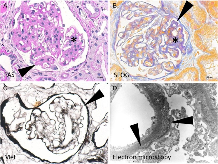
Fig. 2.
Histology of kidney biopsy 2006. (A) Broadened and hypercellular mesangial field (asterisk) as well as focal thickening of peripheral basement membranes (arrowhead, exemplary) is displayed in the PAS staining. (B) The acid fuchsin-Orange G staining displays collagen fibres in blue, which shows double contours of peripheral basement membranes (arrowhead, exemplary) and collagenous matrix in the mesangium (asterisk). (C) Focal double contours are also highlighted in the Methenamine silver stain (arrowhead). (D) Basement membrane lamellation is displayed in the electron microscopy. Endothelial cells and podocytes are slightly autolytic.

