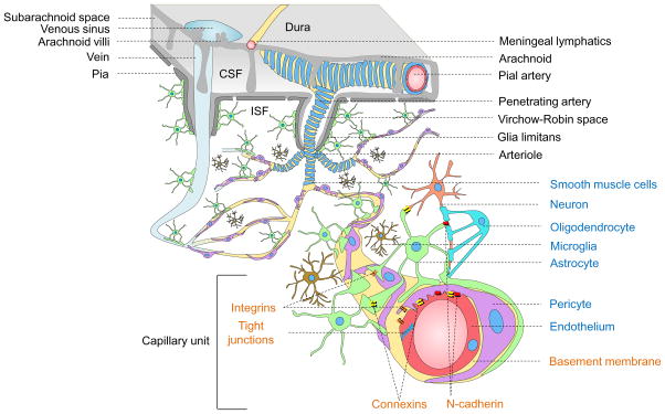
Figure 1. Neurovascular unit.
Vessels in the Subarachnoid space: The subarachnoid space contains cerebrospinal fluid (CSF) that drains via arachnoid villi into the venous sinuses. Cerebral arteries branch into smaller pial arteries. Cerebral veins empty into dural venous sinuses. Meningeal lymphatic vessels carry CSF and immune cells to deep cervical lymph nodes.
Intracerebral vessels: Pial arteries give rise to the penetrating arteries that branch into arterioles all covered by vascular smooth muscle cells (blue). The penetrating arteries are separated from brain parenchyma by the glia limitans, an astrocytic endfeet layer that forms the outer wall of the Virchow-Robin spaces containing brain interstitial fluid (ISF, white). Arterioles branch off into capillaries and the vessels enlarge, as they become venules and veins.
Brain capillary unit: Endothelial cells (red) connected by tight junctions form the blood-brain barrier. Pericytes (purple) and endothelium share a common basement membrane (yellow) and connect with each other with several transmembrane junctional proteins including N-cadherin and connexins. Astrocytes (green) connect with pericytes, endothelial cells and neurons (peach). Microglia (brown) regulate immune responses. Oligodendro-cytes (aqua) support neurons with axonal myelin sheath. Integrins make connections between cellular and matrix components.

