
Figure 5.
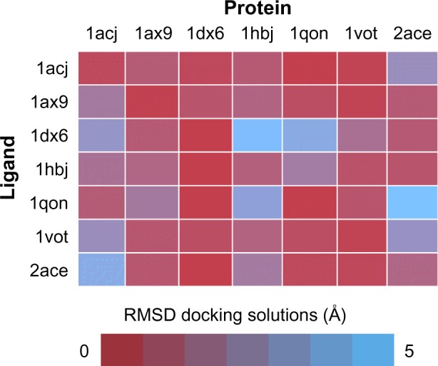
Cross-docking experiment with selected acetylcholinesterase structures from PDB.
Notes: Performance of all possible combinations of rigid docking experiments done in standard conditions, using a series of seven acetylcholinesterase ligands extracted from left column PDB entries onto the same empty protein structures (upper row). Color code indicates RMSD between the best docked solution and the reference PDB.
Abbreviations: PDB, protein data bank; RMSD, root-mean-square deviation.