
Figure 1.
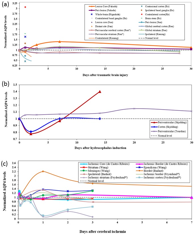
Dynamics of aquaporin-4 (AQP4) expression levels after (A) experimental traumatic brain injury (TBI), (B) hydrocephalus, and (C) cerebral ischemia. (A) Fukuda et al (28) quantified AQP4 levels by immunoreactivity in a rat model of juvenile TBI. Higashida et al (22) measured AQP4 levels in whole-brain lysates with western blotting in a closed-head injury model of rat. Ke et al (26) and Sun et al (24) measured Aqp4 mRNA levels with reverse transcription-polymerase chain reaction in rats. Ren et al (23) measured both global AQP4 levels as well as changes in perivascular AQP4 polarization and found a loss of AQP4 polarization despite a slight global increase. Kiening et al (29) measured AQP4 levels with immunoblotting in ipsilateral and contralateral hemispheres in rats. Subcellular region-specific measurements are denoted with asterisk. Results are shown as fold change compared to the control group. (B) Temporal expression levels of AQP4 after the induction of hydrocephalus in rats. Skjolding et al (37) quantified AQP4 levels over 14 days in the cortex and periventricular zones by western blotting. Using a different model of hydrocephalus, Tourdias et al (38) also observed an elevation of AQP4 in the periventricular region. This increase could have implications in brain water clearance. (C) Temporal dynamics of AQP4 expression levels in the brain after experimental models of ischemia. AQP4 levels after transient focal ischemia were measured by western blotting in mice by de Castro Ribeiro et al (43). The region-specific expression of AQP4 often characterized by an initial increase within a few hours of primary injury was also measured by Wang et al (13) using a transient global ischemia model in piglets. Except in the ischemic core and striatum, AQP4 levels displayed a delayed but significant upregulation 12 to 24 hours after ischemia. Badaut et al (45) quantified AQP4 immunoreactivity in a neonatal rat model of middle cerebral artery occlusion. Frydenlund et al (47) quantified perivascular AQP4 with immunogold labeling in mice subject to 90 minutes of middle cerebral artery occlusion.