
Figure 2.
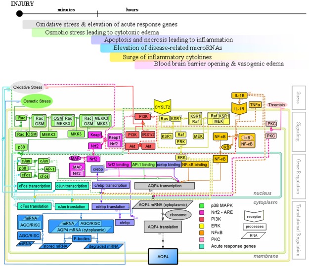
Signaling and regulation of aquaporin-4 expression after ischemic injury. Top frame shows the temporal phases of secondary injury mechanisms following ischemia. The astrocyte cell is divided into three compartments: nucleus, cytoplasm, and membrane. The membrane compartment is designed to illustrate the translocation of signaling molecules towards the membrane. The upper portion of the cell shows signaling events during injury leading to the transcriptional regulation of the aquaporin-4 gene. The center portion shows known transcription factor binding sites on the promoter of aquaporin-4 gene. The lower portion shows translational regulation events by microRNAs. The extracellular insults above the cell are arranged according to the approximate timeline of secondary injuries after cerebral ischemia. “P” within a circle marking the upper-right corner of certain molecules represents the phosphorylated state, and dashed lines indicate that the detailed signaling mechanism is still unknown. Arrows indicate activation, while flat caps indicate inhibition. The figure is generated in CellDesigner (153).